Temporal Bone Imaging
Nonfiction, Health & Well Being, Medical, Specialties, Radiology & Nuclear Medicine| Author: | ISBN: | 9781588906472 | |
| Publisher: | Thieme | Publication: | January 1, 2011 |
| Imprint: | Thieme | Language: | English |
| Author: | |
| ISBN: | 9781588906472 |
| Publisher: | Thieme |
| Publication: | January 1, 2011 |
| Imprint: | Thieme |
| Language: | English |
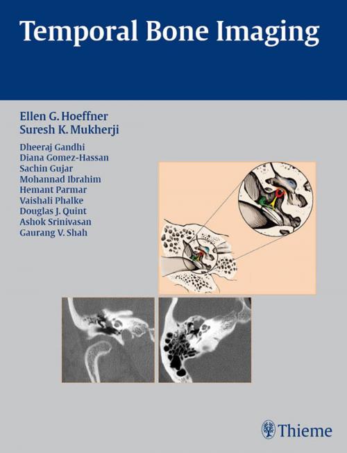
Temporal Bone Imaging is a case-based review of the current techniques for imaging the various temporal bone pathologies frequently encountered in the clinical setting. Detailed discussion of anatomy provides essential background on the complex structure of the temporal bone, as well as the external auditory canal, middle ear and mastoid air cells, facial nerve, and inner ear. Chapters are divided into separate sections based on the anatomic location of the problem, with each chapter addressing a different disease entity.Highlights:Each chapter features succinct descriptions of epidemiology, clinical features, pathology, treatment, and imaging findings for CT and MRI Bulleted lists of pearls highlight important imaging considerations More than 200 high-quality images demonstrate anatomy, pathologic concepts, as well as postoperative outcomes This book will serve as a valuable reference and refresher for radiologists, neuroradiologists, otologists, and head and neck surgeons. Its concise, case-based presentation will help residents and fellows in radiology and otolaryngology-head and neck surgery prepare for board examinations.
Temporal Bone Imaging is a case-based review of the current techniques for imaging the various temporal bone pathologies frequently encountered in the clinical setting. Detailed discussion of anatomy provides essential background on the complex structure of the temporal bone, as well as the external auditory canal, middle ear and mastoid air cells, facial nerve, and inner ear. Chapters are divided into separate sections based on the anatomic location of the problem, with each chapter addressing a different disease entity.Highlights:Each chapter features succinct descriptions of epidemiology, clinical features, pathology, treatment, and imaging findings for CT and MRI Bulleted lists of pearls highlight important imaging considerations More than 200 high-quality images demonstrate anatomy, pathologic concepts, as well as postoperative outcomes This book will serve as a valuable reference and refresher for radiologists, neuroradiologists, otologists, and head and neck surgeons. Its concise, case-based presentation will help residents and fellows in radiology and otolaryngology-head and neck surgery prepare for board examinations.
